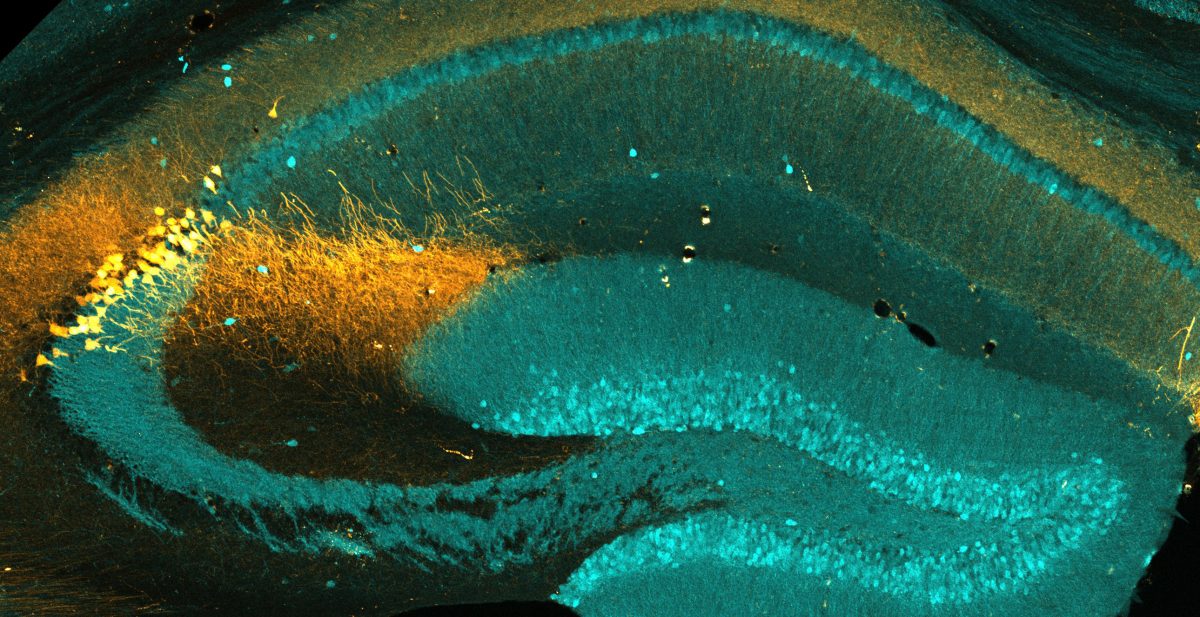
Amigo2-expressing CA2 neurons were genetically engineered to express Cre-recombinase, which was then used to express a red fluorescent reporter protein (tdTomato). Neighboring CA1 and Dentate Gyrus neurons are stained with an antibody against calbindin 1 (pseudo-colored in cyan). This tissue was physically expanded nearly 100-fold in volume prior to imaging using a technique called Expansion Microscopy, which was developed in 2015 by the Boyden lab at MIT (Chen, Tillberg & Boyden, Science, 2015). This technique allows for dense, compact tissues, such as in the brain, to be physically separated and optically resolved to visualize the finer details of neuronal architecture.

