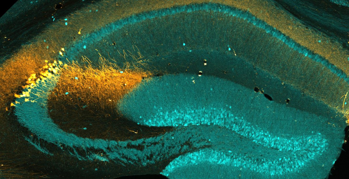
A 60X confocal image of hippocampal CA2 neurons labeled with CA2 marker Necab2 (magenta), mitochondrial marker PDH (cyan) and sparsely labeled Tdtomato (pseudocolored blue). This brain section was expanded ~4.5x in volume (note the scale bar is 50 microns) using expansion microscopy to better visualize subcellular morphology.
